Объявления
-
Памятки «Навигатор профилактики», разработанные Московским...
Уважаемые преподаватели и кураторы ФГБОУ ВО ДГМУ Минздрава России! В дополнение к ранее направленному письму от 13 октября 2022 г. № 06-14207/09-18/22 и в...
13.12.2022
2040
-
Список научных и педагогических сотрудников, относящихся к...
Список ППС
02.07.2024
21
-
Как вести себя при обстреле
25.06.2024
78
-
Что делать при угрозе ракетной атаки или обстрела...
25.06.2024
69
-
Новые учебные планы 2024 г.
Лечебный факультет Учебный план 2024-2030 г. Учебный план 2023-2029 г. Учебный план 2022-2028 г. Учебный план 2021-2027 г. Учебный план 2020-2026 г. Учебный план 2019-2025...
25.06.2024
1367
-
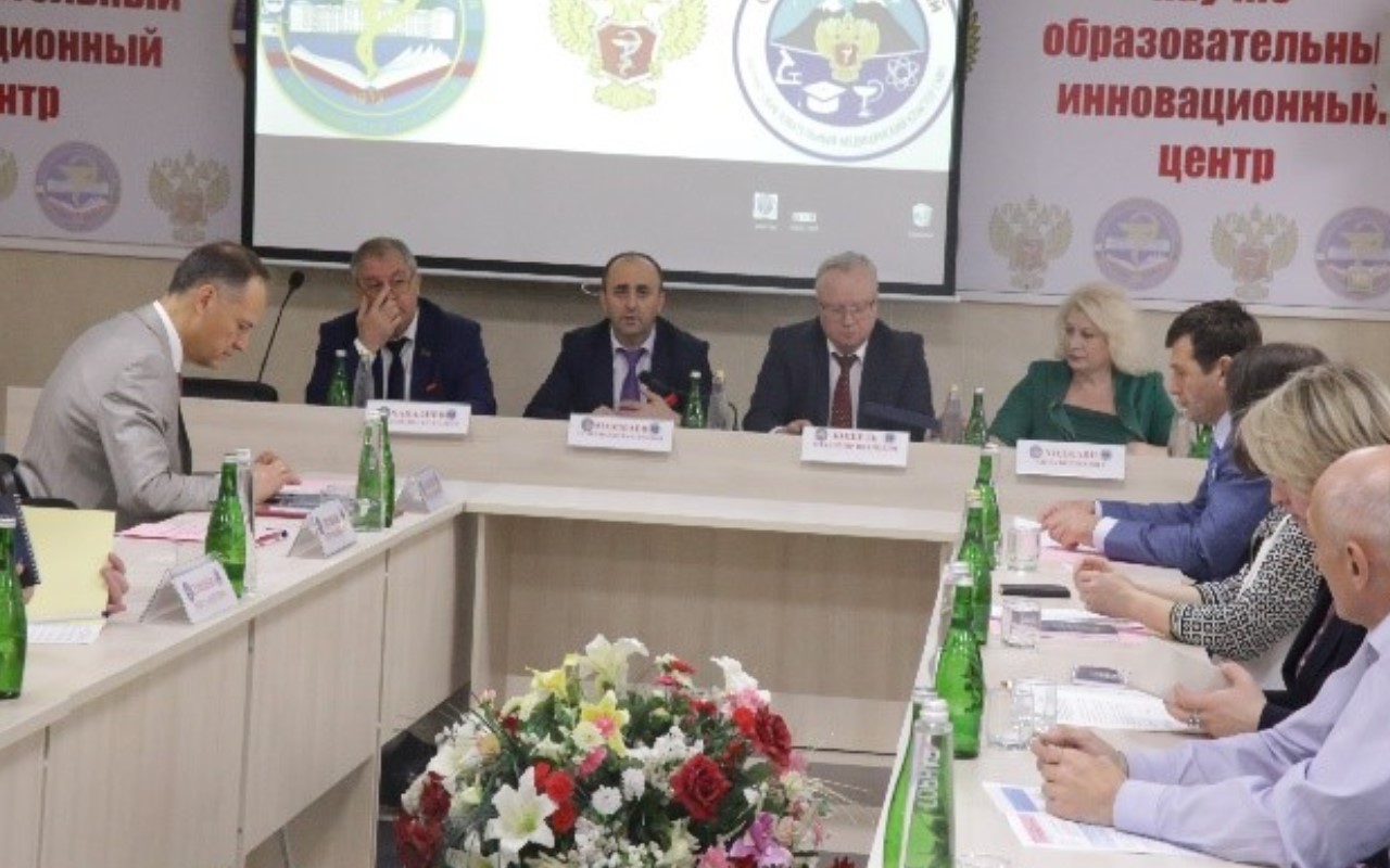
Очередное заседание Ученого совета
Уважаемые члены Ученого совета! 28 июня 2024 года в 16.00 состоится очередное заседание Ученого совета на втором этаже в зале заседания (Пл. Ленина, 1).
21.06.2024
185
-
Дайджест возможностей для молодых ученых
Продолжается прием заявок на конкурсы, гранты для поддержки молодых ученых, ниже представлены ссылки на активные конкурсы: Перечень
21.06.2024
69
-
Телефон для приема сообщений о коррупционных проявлениях:
68-32-80
-
Эл. почтовый ящик:
Ubdgmu@mail.ru

Department of Medical Simulation and Educational Practice
11.06.2021
284
Head of the department: Koichuyev Rasul Abaakarovich
General information
Address: Makhachkala, Dagestan State Medical University, I. Shamil av., 46.
E-mail: sim.center_dgmu@mail.ru
Telephone: 8929 875 30 86
In 2018, at the suggestion of Rector S. N. Mamaev, the Academic Council of the University considered positive the issue of creating a department of medical simulation and educational practice on the basis of a training simulation center.
LOCATION
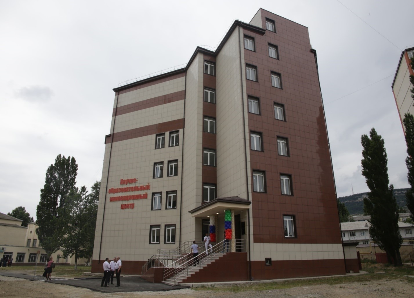
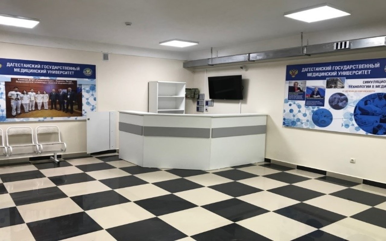
The Department of Medical Simulation and Teaching Practice is located in the new modern seven-storey building of the University at Shamil 44.
The department is deployed in two academic blocks: the first block occupies the area of premises in the educational and laboratory building of the University at Shamil 46 (total area of 500m2), as well as a separate block in the new modern building of the University on two floors, with a total area of 1200 m2.
DEPARTMENT OF MEDICAL SIMULATION AND TRAINING PRACTICE
Purpose of the department creation
Improving the quality of practical training of students: students, residents, medical cadets in professional retraining and advanced training programs through the use of modern and simulation technologies for mastering and improving practical skills-simulators, phantoms, as well as virtual (computer) simulators that provide high realism of medical interventions and procedures.
On the basis of the Department of Medical Simulation and Training Practice, a consistent continuity-transient model of the learning process for mastering, training and strengthening practical skills is implemented. Simulation technologies are conducted in the following areas of training: therapy, cardiology, obstetrics-gynecology, anesthesiology and resuscitation, surgery, including endoscopy and laparoscopy (virtual simulation technologies). It is envisaged to master basic general medical and special skills.
Objectives: Effective and high-quality training in professional competences in accordance with federal state educational standards, developing practical skills for students on phantoms, simulators, simulators in accordance with the requirements of professional standards and preparing students for accreditation and conducting a specialist accreditation procedure were the main tasks of the department, which are dictated by the current state of medical education issues.
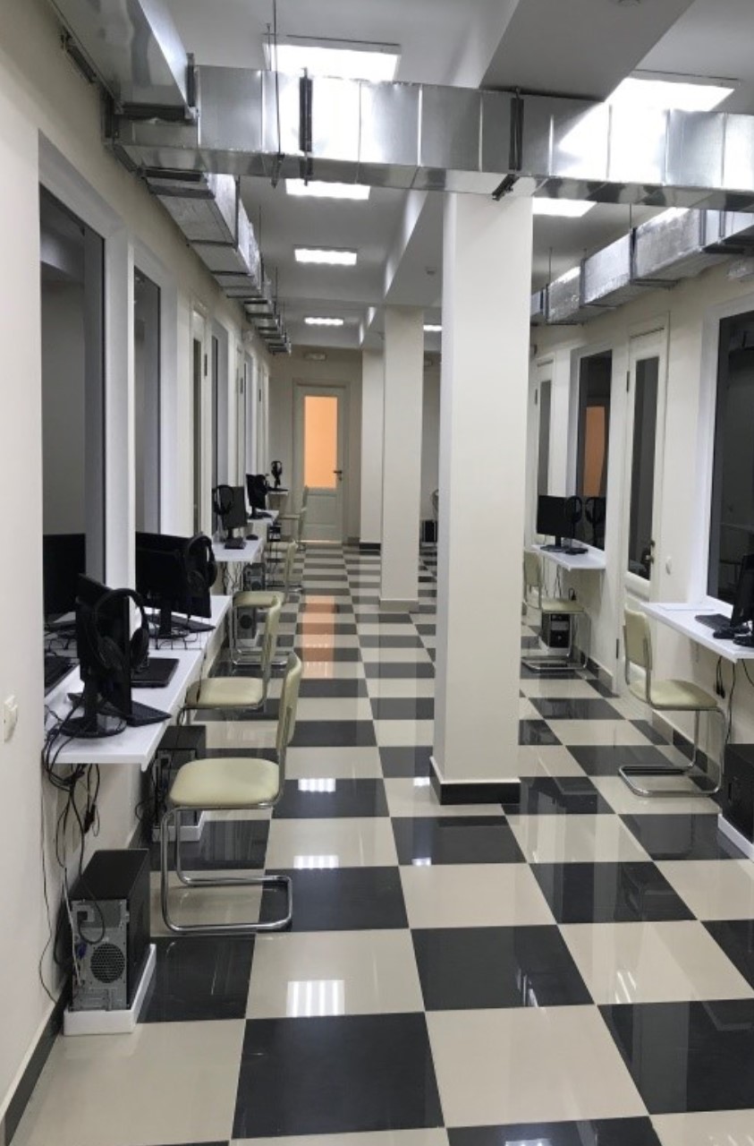
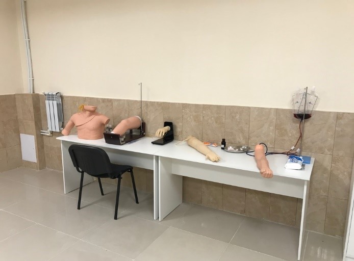
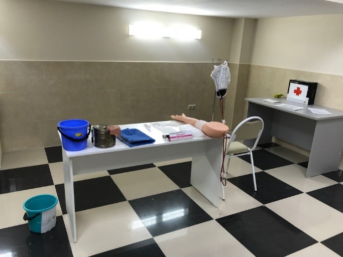
The Department arrangement:
— classrooms;
— stations;
— skill training hall in anesthesiology and resuscitation;
— general medical practice hall with simulator mannikins;
— perinatal hall;
— halls for the manual skills development in endoscopic surgery and the practice of laparoscopic surgery techniques;
— General Surgery Hall;
— procedure rooms;
— operating room.
Staff:
- Head of the department: Koichuyev Rasul Abaakarovich.
- Head of the teaching department:assistant Abdurazakova Milana Abdurazakovna
• Candidate of Medical Science, Associate Professor Aliyev Basir Omarovich
• Candidate of Medical Science, Associate Professor Aselderova Aida Shamsutdinovna
• Candidate of Medical Science, assistant Omarova Patimat Magomedovna
• Candidate of Medical Science, assistant Magomedova Patimat Aripovna
• Candidate of Medical Science, assistant Abserkhanova Javryat Ubayteyevna
• Candidate of Medical Science, assistant Kurbanismayilova Rahimat Ramazanovna
• Assistant Abdullayeva Patimat Kamilievna
• Assistant Suleymanova Khazina Magomedovna
• Senior laboratory assistant Aisayeva Bahu Magomedovna
The department is equipped with advanced high-tech equipment, allowing to conduct the training process with the help of simulation technologies at the highest and most modern level, meeting the world requirements of specialists training. The department training facilities presented by mannequins, dummy models, medical simulators, simulators, highly realistic robots.
“By three methods we may learn wisdom: First, by reflection, which is noblest; Second, by imitation, which is easiest; and third by experience, which is the bitterest.”
― Confucious (V century BC.)
«Simulation» – a person, a device, efforts to recreate the problem, where the student shall react as he will do in a real situation».
Traditional educational system in its current expression, it is based primarily on knowledge, while in a professional environment a specialist is assessed according to the criteria of skills and abilities. An important advantage of the use of simulation technologies in the educational process is the ability to objectively register the parameters of the doctor’s activities in the aspect of the developed professional skills and abilities. The training task is to process algorithms for emergency care and intensive care in a limited period of time (seconds-minutes). In the simplest form of simulation can be described as an imitation of reality. In the medical education system, simulations are the basis of a number of techniques designed to reproduce clinical situations for the purpose of learning, repetition, evaluation, and research. Simulators range from simple physical models of anatomical structures, to complex devices and mannequins with high mechanical reality and computer control. Simulation technologies allow simulate various clinical situations, including rare clinical scenarios.
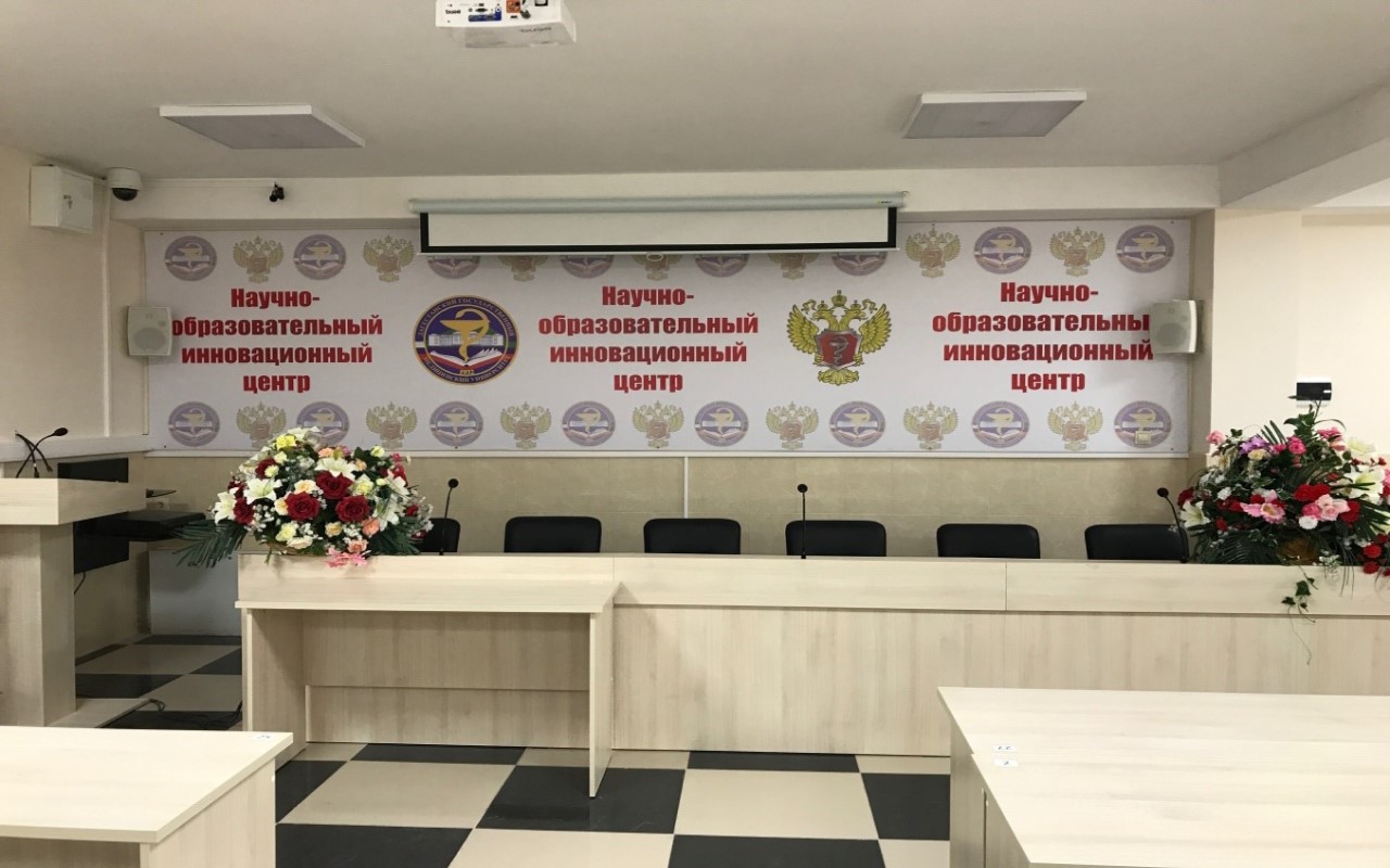
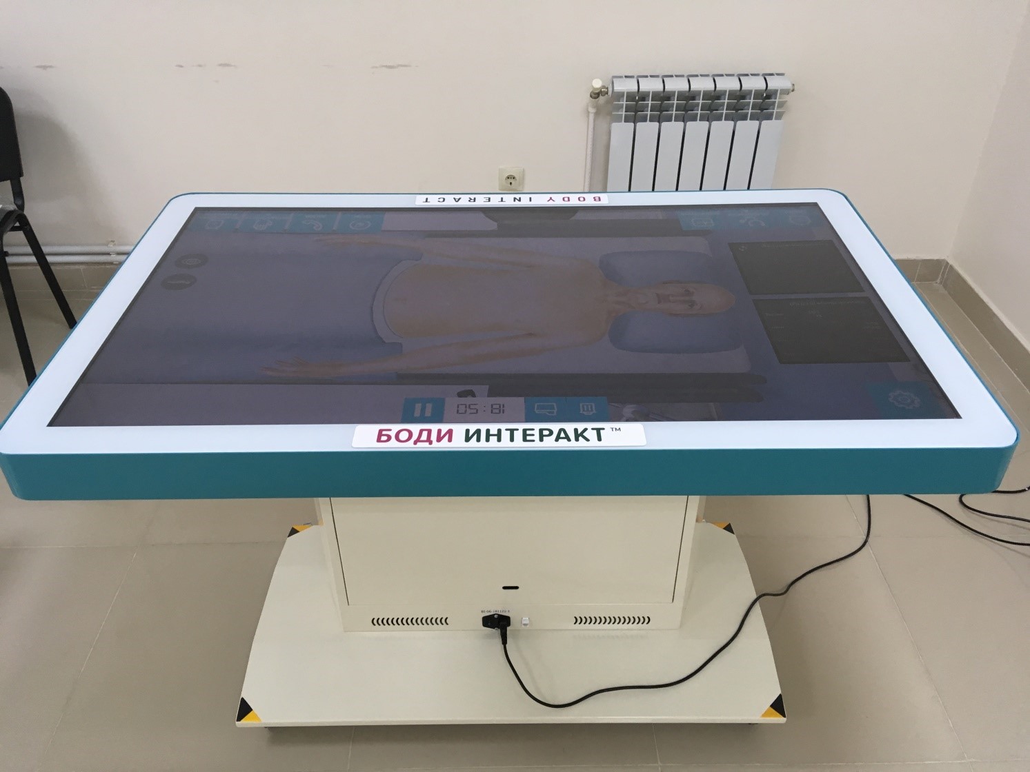
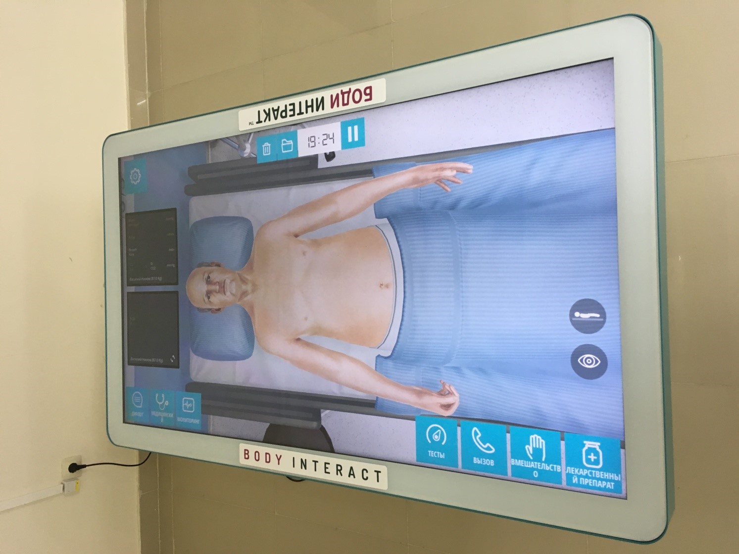
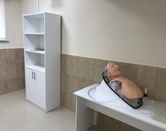
Medical Simulation and Accreditation Center Department facilities


Conference hall
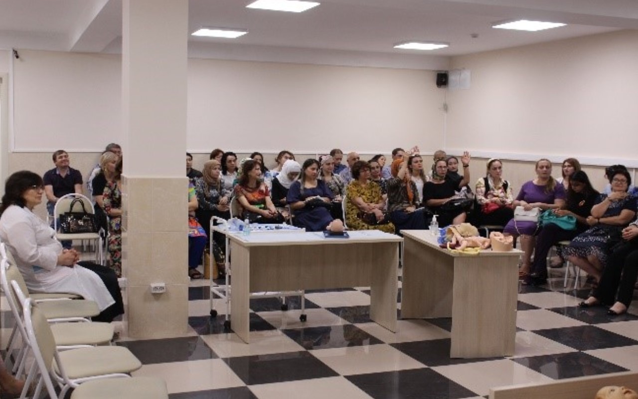


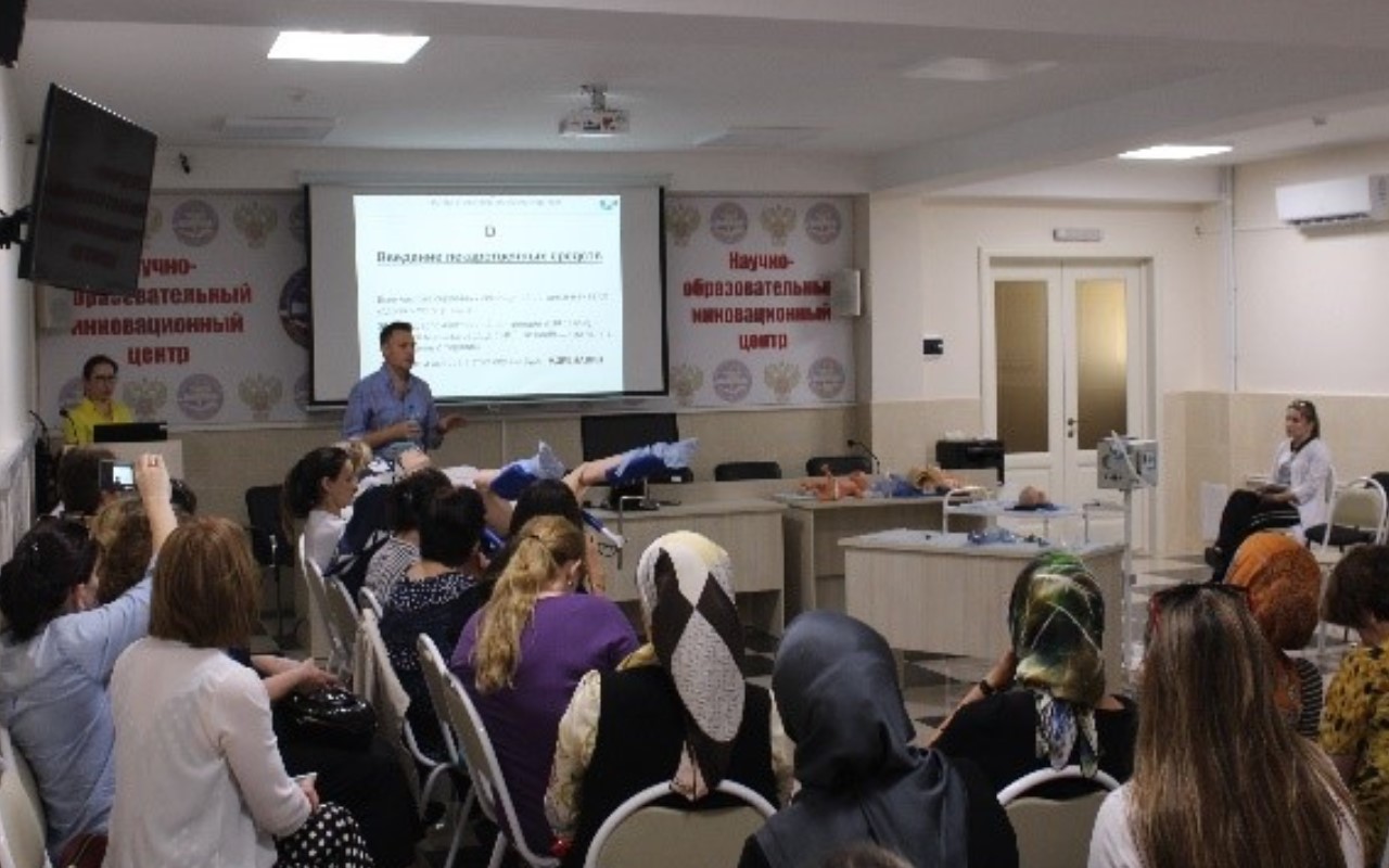
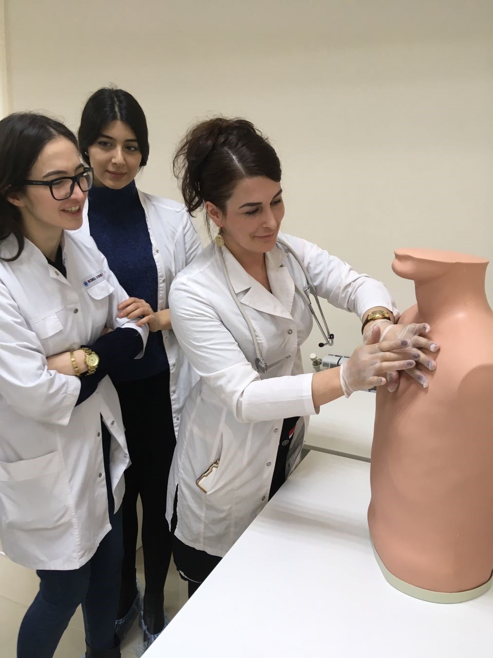
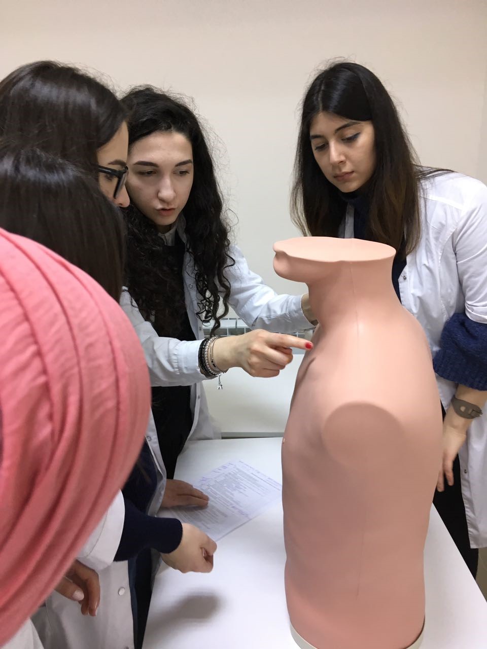
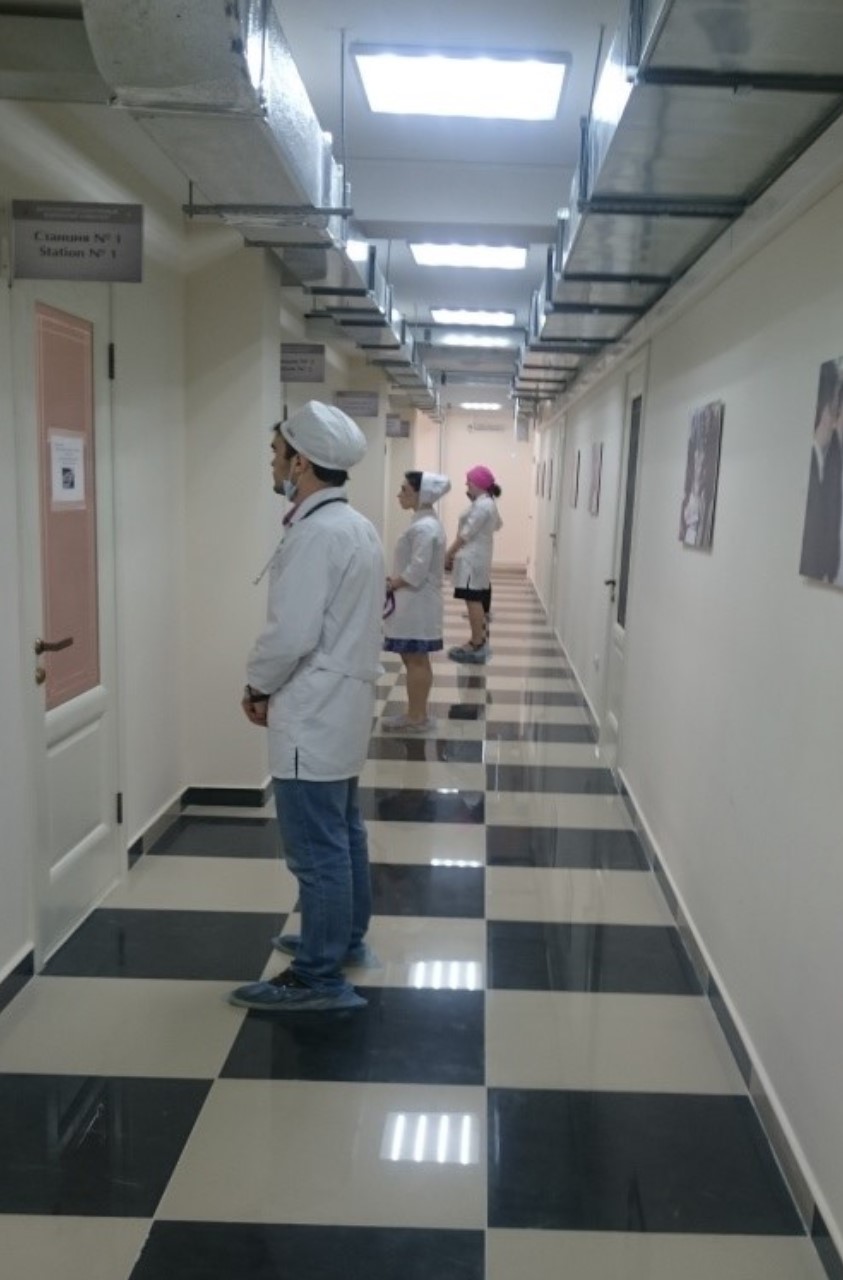
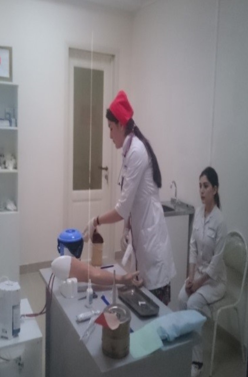
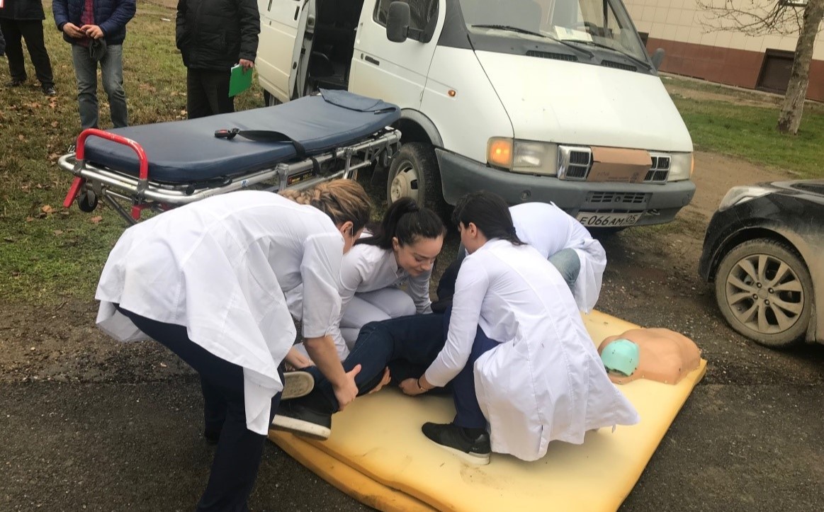
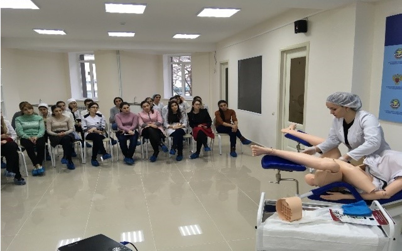
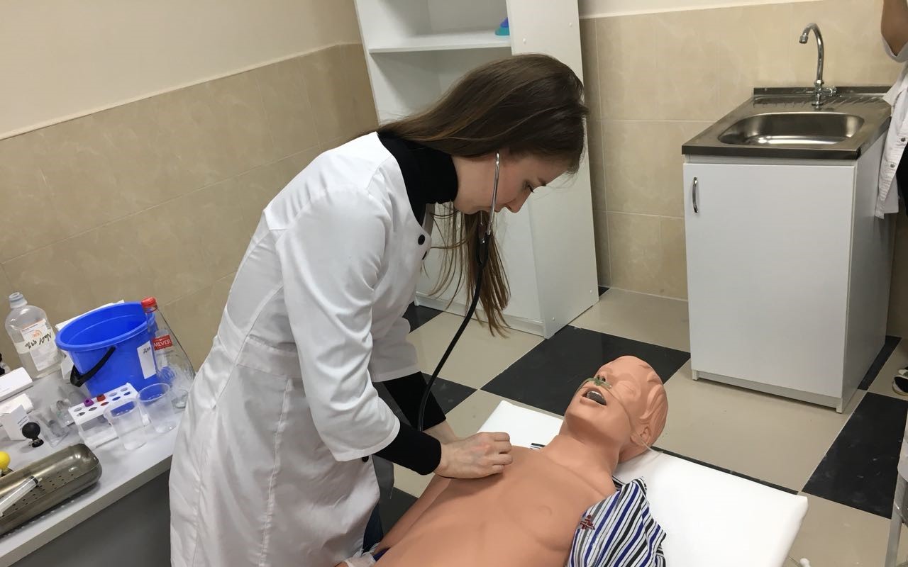
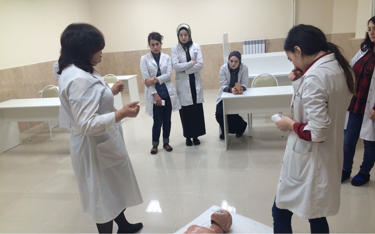
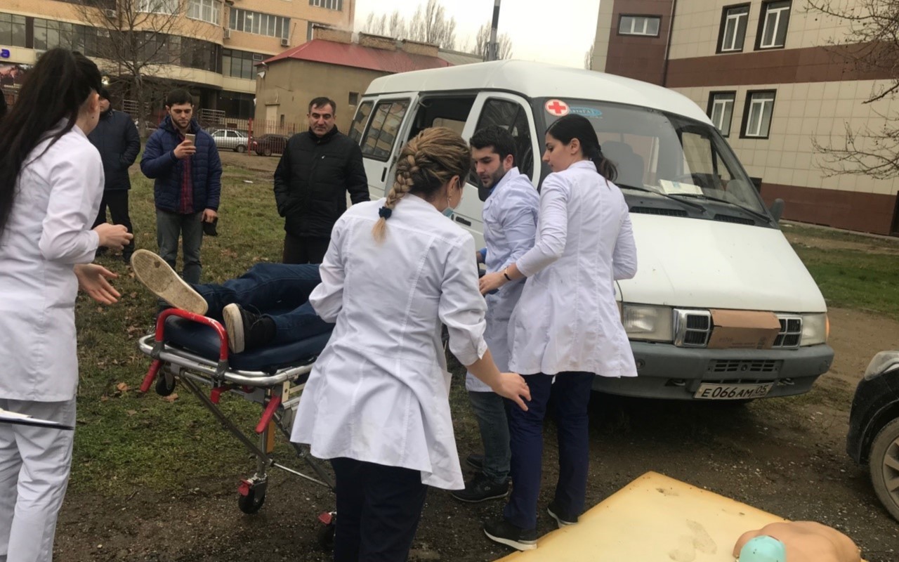
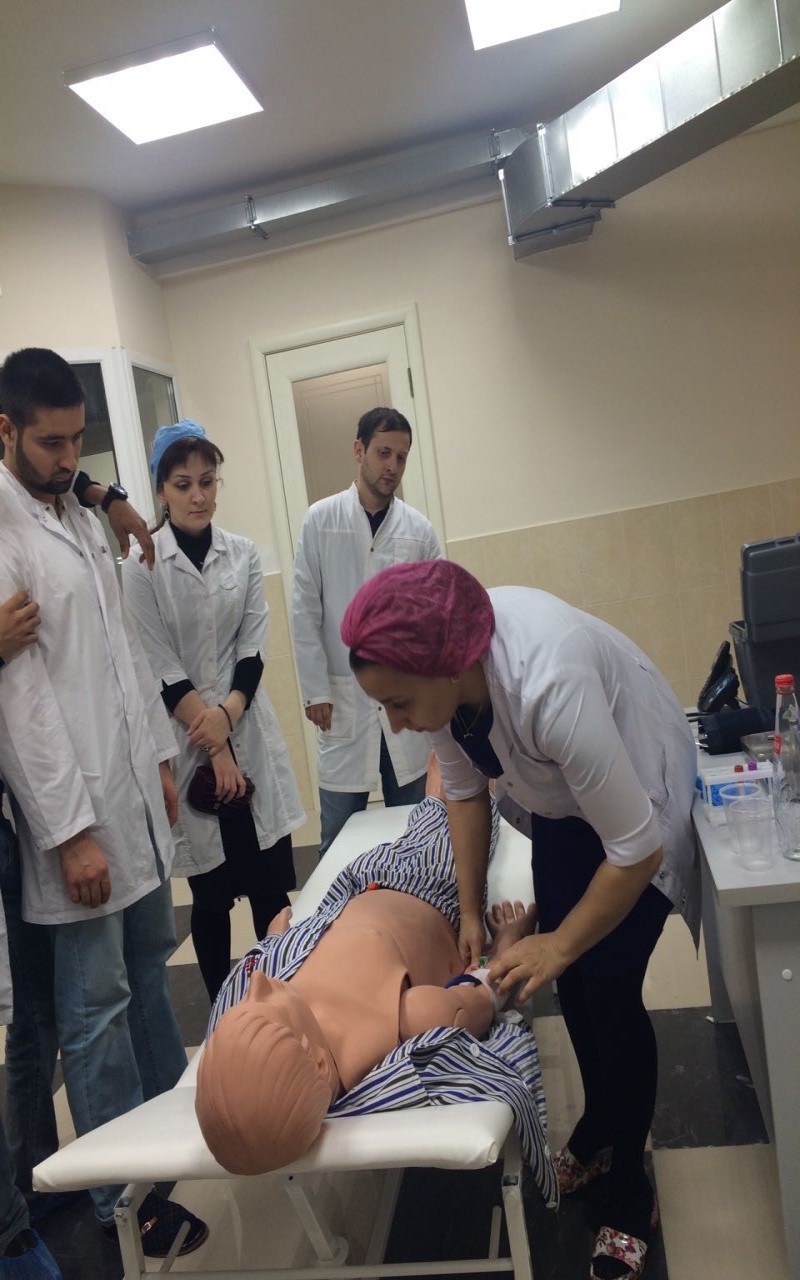
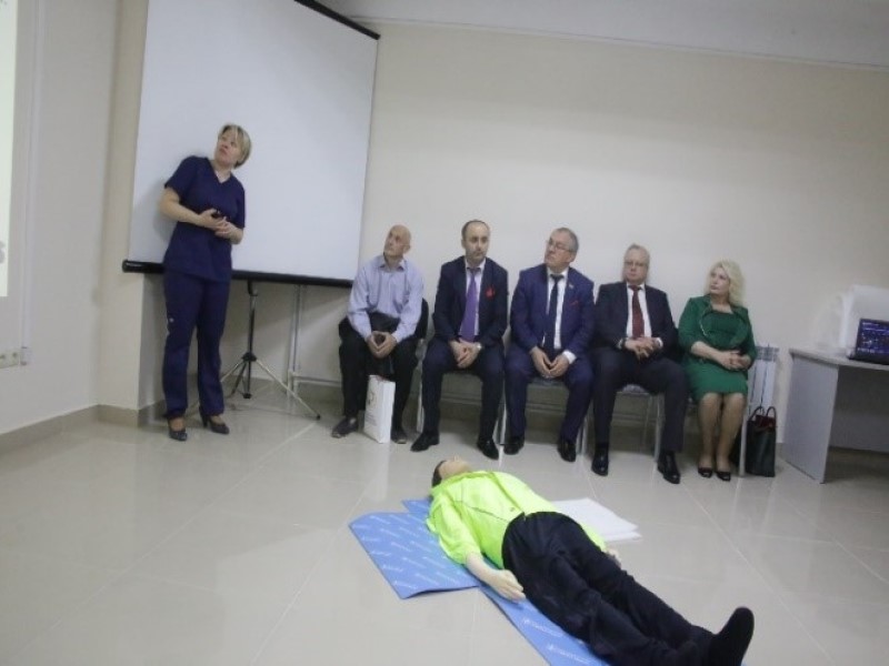
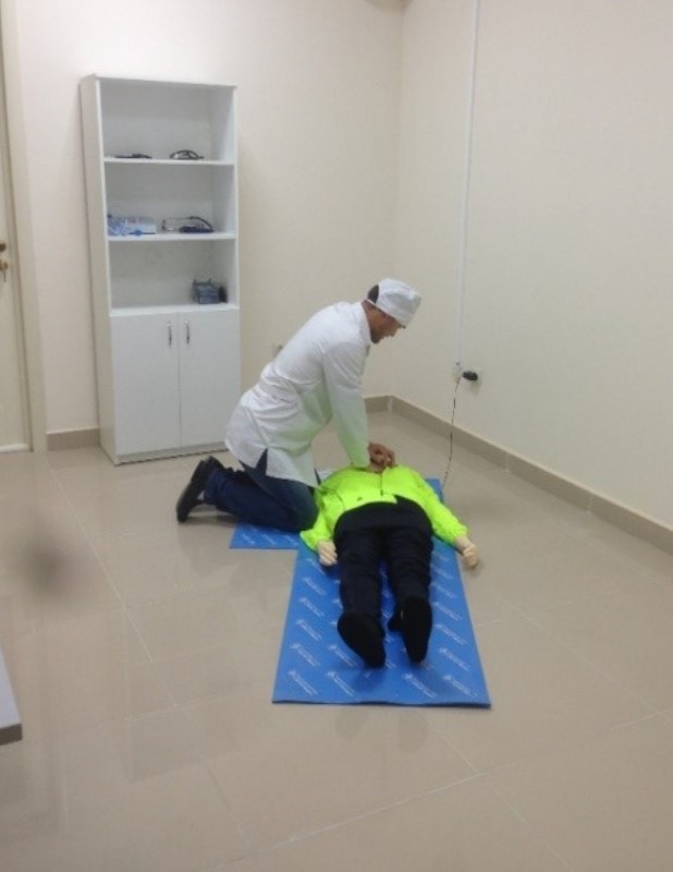
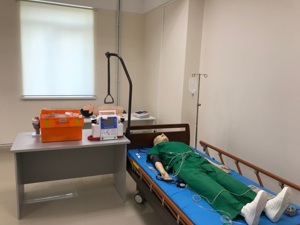
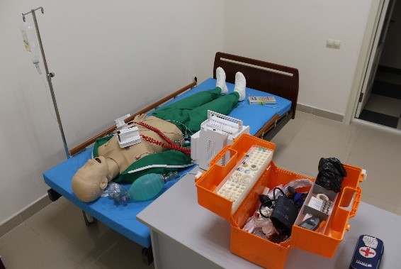
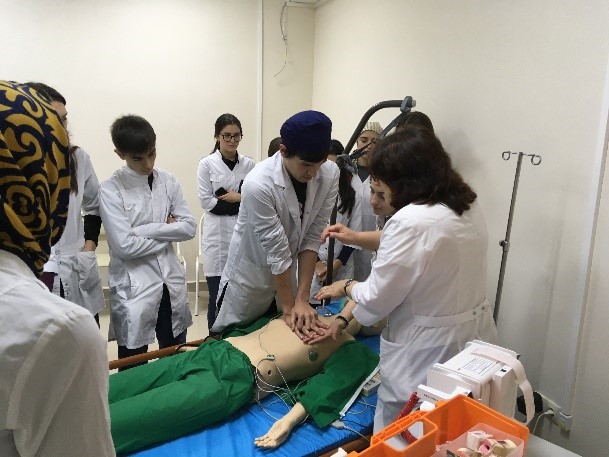
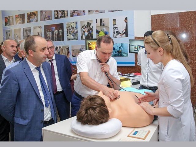
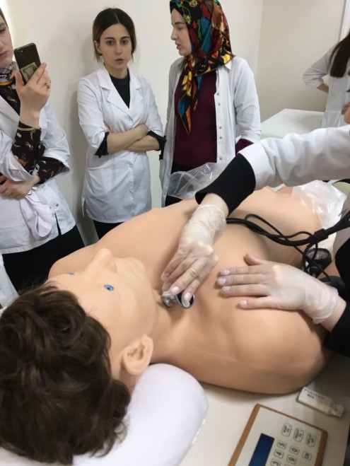
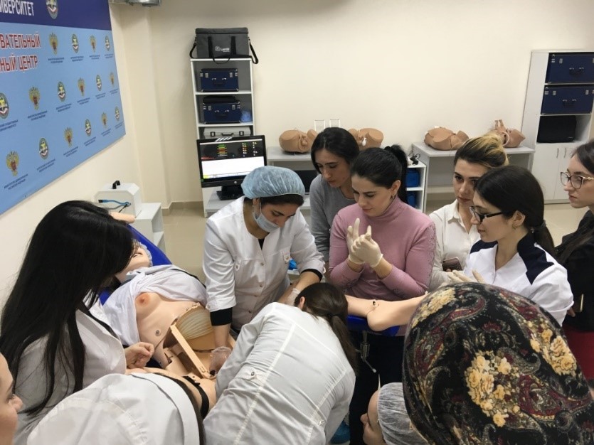
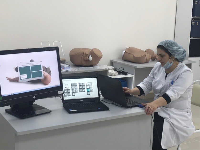
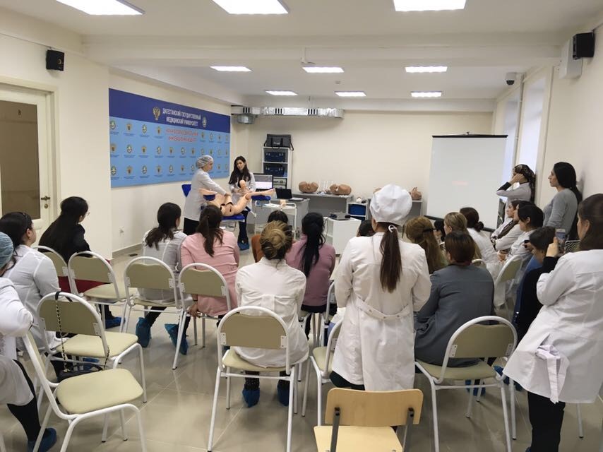
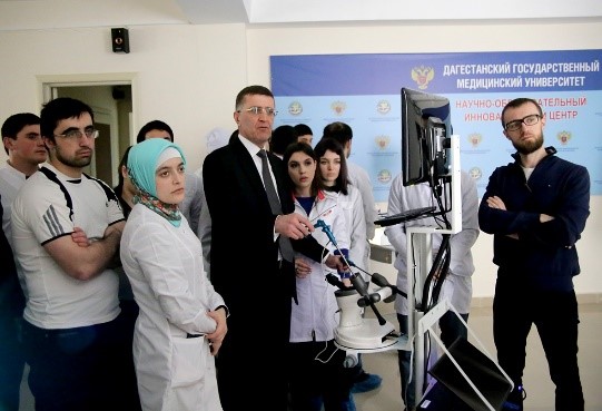
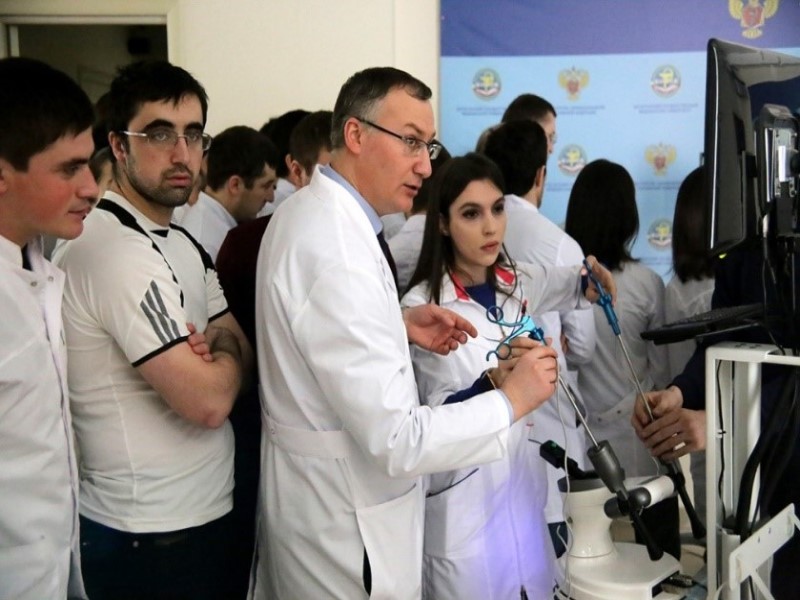
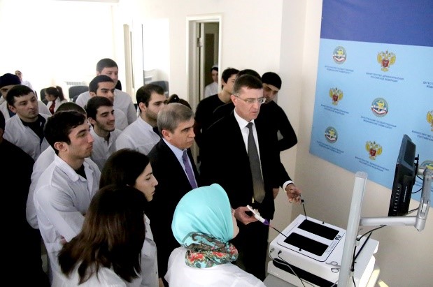
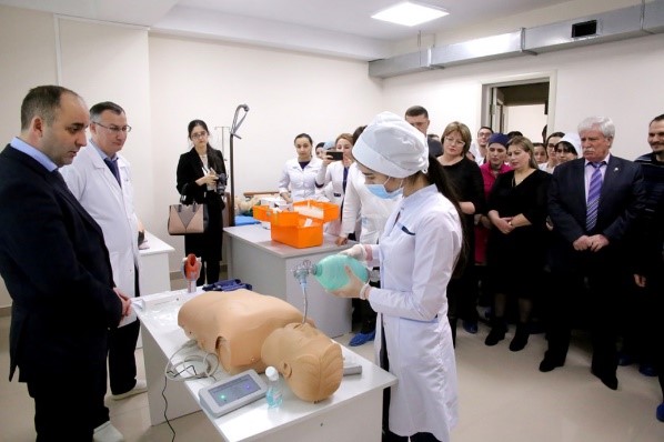
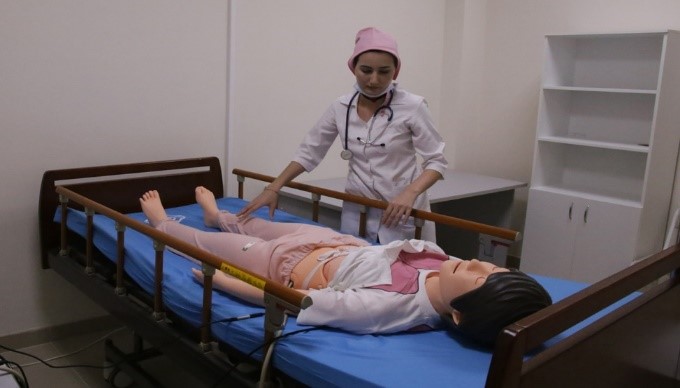
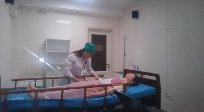
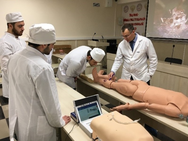
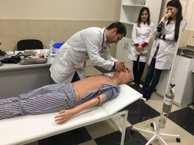
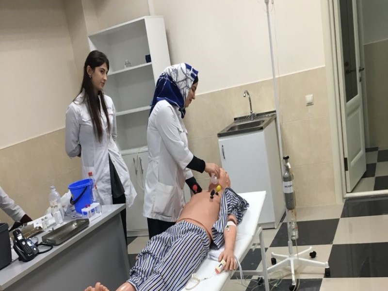
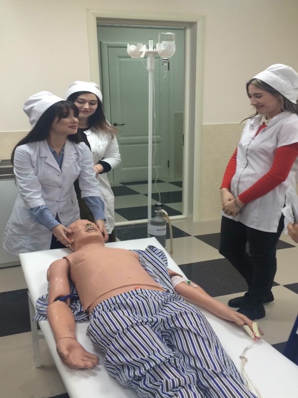
Accreditation center and department of medical simulation and training practice are equipped with the latest tools and technologies of training: simulators and robots, simulation mannequins, electronic phantoms, models and other interactive computerized equipment, as well as real medical equipment (ECG, video laringoscope, surgical and obstetric instruments, stethoscopes, tonometers, etc.). On the basis of the Department of Medical Simulation, the training of faculty, students, residents, cadets on the programs of the cycle of professional development and professional retraining with the use of simulation technologies is carried out; Since 2016, the procedure of accreditation of DSMU graduate specialists is taking place. At the Department of Medical Simulation and Educational Practice, practical classes for students of 2-6 courses of medical and pediatric faculties are regularly organized in a systematic manner. At the stage of pre-practical training, students are given the opportunity to work out the skills provided for in the practice program on simulators and simulators, and get the advice of a teacher. Students are also provided with comprehensive methodological support: implementation algorithms, video tutorials and expert assessment sheets for all practical skills are developed and posted on the official website of the university. At the Department of Medical Simulation and Training Practice, to increase the objectivity of evaluating the correctness of performing practical skills on training dummies, the test / exam is conducted using video monitoring on expert assessment sheets (checklists). The teacher, being in another room, evaluates the correctness of the implementation of practical skills by students on a computer monitor, simultaneously filling out sheets of expert assessments, which note compliance with the sequence and correctness of the implementation of the practical skill algorithm. At the Department of Medical Simulation and Educational Practice of DSMU, all practical classes are conducted using debriefing technology, when the process of learning and practicing practical skills is recorded on video cameras, and then broadcast to the debriefing hall for detailed analysis and analysis of students’ mistakes. The Department of Medical Simulation and Educational Practice regularly organizes various competitions, Olympiads, trainings, and master classes. The Practical Skills Competition encourages students to improve their theoretical knowledge and practical skills in providing emergency and emergency emergency medical care; contributes to the development of students’ creativity and interest in the practical activities of the doctor.
AT THE DEPARTMENT OF MEDICAL SIMULATION AND EDUCATIONAL PRACTICE, TRAINING IS CONDUCTED IN SPECIALTIES:
- Nursing, care for sick adults and children
• Premedical first aid
• Emergency care, resuscitation, anesthesiology
• Therapy
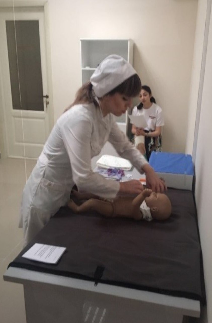
• Pediatrics and neonatology
• Internal diseases
• Obstetrics and Gynecology
• Surgery
• Traumatology and Orthopedics
• Neurology and neurosurgery
• Dentistry


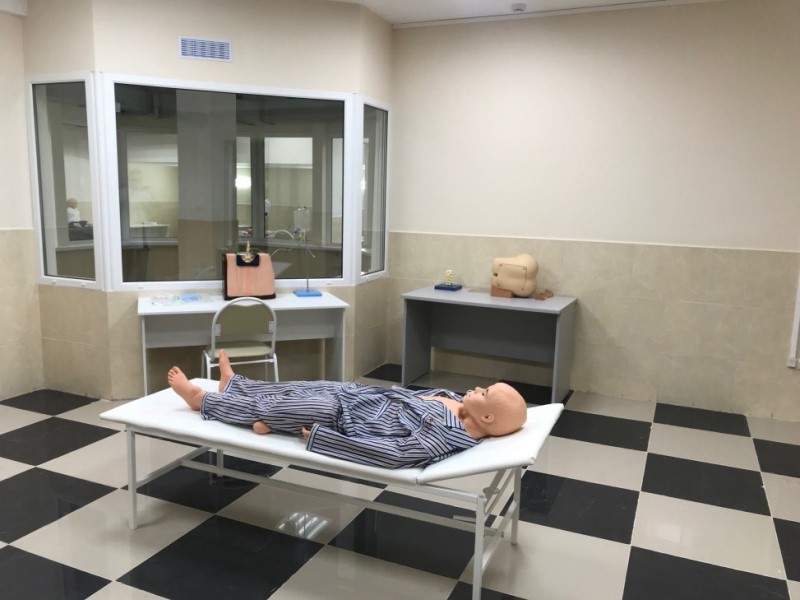
THE DEPARTMENT ARRANGEMENT OF MEDICAL SIMULATION AND TRAINING PRACTICE INCLUDES:
Classrooms, simulation rooms: emergency and resuscitation unit for adults and newborns, obstetric and gynecological manual unit, traumatology and surgery unit, therapy unit, cardiology unit, neurology unit, dentistry unit, pharmacy unit (training pharmacy), teaching and technical staff offices.
Simulation equipment and equipment of the Department of Medical Simulation and Educational Practice and Accreditation Center:
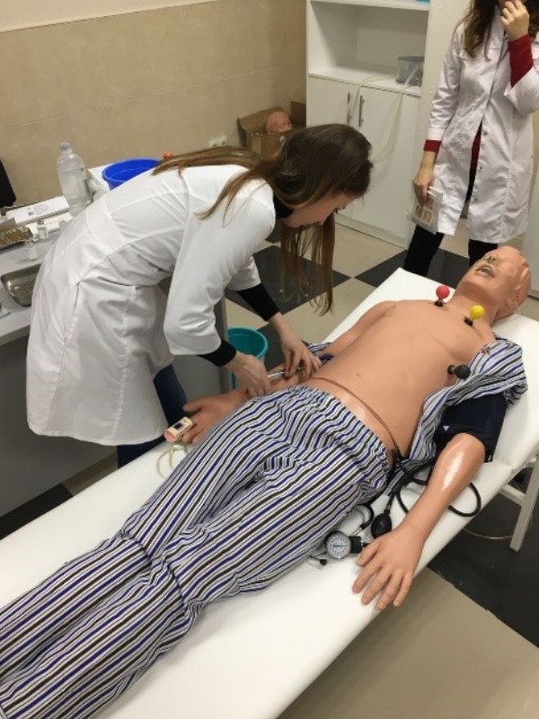
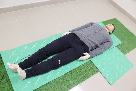 RODAM. An adult resuscitation simulator with wireless control for providing emergency care in a team for various conditions, with the ability to monitor and record basic vital signs (Korea).
SKILLS
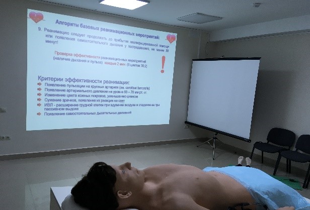
- performing cardiopulmonary resuscitation
- carotid pulse determination
- respiratory tract patency determination
- automated external defibrillator
- determining the level of consciousness
SPECIFICATIONS
- the simulator allows you to practice the skills of providing emergency care in a team in various conditions with the ability to monitor and record basic vital signs
- self-guided breathing with chest excursion, carotid artery pulsation, pupillary reflex
- wireless connection to the computer
- simultaneous connection of several simulators to a single laptop
- D-type battery connection
- maximum chest compression depth — 7 см
- when compressing the chest, the depth of compression and the correct hand position are displayed in real time on the laptop screen
- with artificial ventilation, the exhalation rate and the volume of ventilation are displayed in real time
- during artificial ventilation, the sensor measures the volume of air and automatically adjusts the chest excursion
- ensuring the patency of the respiratory tract, the degree of head tilt is checked (registration of hyperextension)
- power mode is selected by the simulator automatically
- the ability to select and customize various recommendations for CPR and training and evaluation regimes
- the ability to set criteria for the assessment regime
- the ability to change the state of the simulator in real time in training mode
- the ability to save data for analysis and debriefing, print reports
- the possibility of creating groups of students, analyzing averages by group, etc.

Advanced emergency care simulator Saveman

SKILLS
• endotracheal intubation
• artificial respiration with a laryngeal mask and a combination of breathing tubes
• oral, nasopharyngeal intubation
• diagnosis of unilateral lung intubation by chest augmentation
• lungs breathing listening by direct auscultation with a stethoscope over the clavicle and in the axillary region
• diagnosis of air ingestion in the stomach
• infusion therapy
SPECIFICATIONS
• the simulator presents options for abnormal heart rate and ECG, you can choose 16 main and 4 additional options for ECG rhythm
• the ECG rhythm can be changed by remote control
• the remote control can operate wirelessly on battery power
• the common carotid artery is palpated, its pulsation is synchronized with the ECG rhythm, it can be stopped, it is possible to simulate pulse-free electrical activity, ventricular tachycardia and pulse-free ventricular tachycardia
• the ECG rhythm can be transformed after defibrillation and percutaneous pacing, it is possible to set a threshold value for percutaneous pacing
• the ECG rhythm, caused by chest compression, can be incorporated into the main ECG rhythm at a compression depth of more than 4 cm
• ECG waves can be simulated, and defibrillation functions are automatically activated
• up to 20 types of ECG waves can be stored in the simulator’s memory for each scenario
SAM II – auscultatory dummy
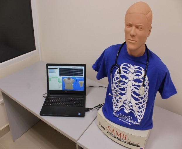

SAM II, auscultatory mannequin, is an interactive dummy for teaching students the art of ausculation. THE SAM provides four areas of listening to heart sounds, eight areas of listening to the sounds of breathing, two areas of listening to bowel sounds and one area to determine the pulse of the carotid artery. The sound library has 16 cardio/pulmonary combinations, 27 heart sounds, 21 sound of breathing and 20 bowel sounds.
 The areas of listening to heart sounds are located in the areas of the valves (aortic, tricuspid, pulmonary artery and mitral) on the chest, and the areas of listening to breathing sounds are divided into anterior (left upper and lower; right upper and lower) and posterior (left upper and lower; right upper and lower). The areas of listening to the sounds of the intestine are located in the upper, right and left squares.
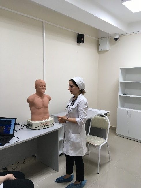
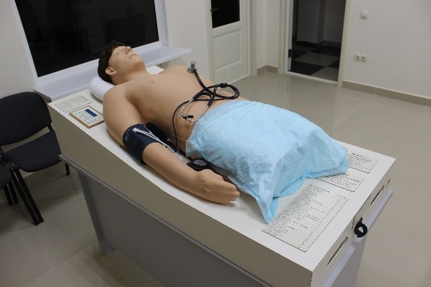
A simulator for the physical examination of a Harvey cardiology patient.
SKILLS
Physical examination of cardiac patient
SPECIFICATIONS
• the simulator is an adult male mannequin, mounted on a stand in a horizontal position
• the dummy has a head with a realistic-looking scalp, open eyes, torso, upper limbs, and lower limbs up to the upper third of the thighs
• the mannequin body reproduces all the anatomical landmarks characteristic of the reproduced parts of the human body
• the simulator reproduces the physical data of various heart diseases and allows you to measure the following parameters:
— blood pressure on the right hand
— frequency and pulse filling on jugular veins
— pulsation on the carotid arteries – on both sides
— pulsation on the shoulder artery – on the right side
— pulsation on the femoral arteries on both sides
• the pulse wave shape corresponds to a reproducible disease
• simulator allows you to listen to the precardial pulsation at the following points:
— pulmonary artery valve kidney (left upper edge of the sternum)
— right ventricle (middle or lower left edge of the sternum)
— left ventricle (apex -5th intercostal space, midclavicular line)
apical beat displacement (6th and 7th intercostal space)
— anterior axillary line
• different types of precardial pulsation are programmed for each reproducible disease
• the following areas for heart ausculation are presented:
— aortic valve listening point (the upper right edge of the sternum)
— listening point of the pulmonary artery valve (the upper left edge of the sternum)
— listening point of the tricuspid valve (the left bottom edge of the sternum)
— mitral valve listening point (apex)
— additional listening point for the aortic valve or pulmonary artery valve (upper chest area)
— carotid arteries
• the following areas for lung ausculation are presented:
—upper right
—right inferoposterior
—right lower frontal
— upper left
— left lower frontal
— left inferoposterior
• the sounds reproduced during auscultation of the lungs correspond to the selected heart disease
- simulator reproduces the abdominal type of breathing
• all areas of pulsation and sound phenomena are synchronized
• the nature of cardiac noise varies depending on the frequency of breathing
• the built-in speaker has a system for connecting to a radio microphone
• the wireless radio microphone can be attached to the buttonhole
• for each reproducible pathology, different types of blood pressure, precordial pulsation, and heart and lung murmurs are provided
• the simulator has more than 30 training programs
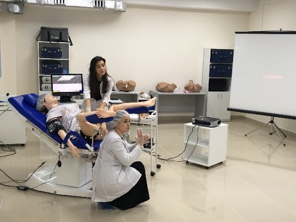
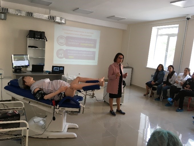
SimMom Childbirth Simulator
SimMom device overview
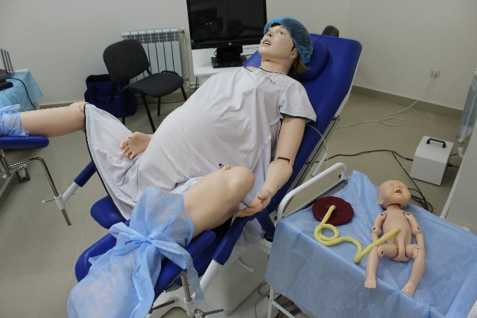 The SimMom device was developed by Laerdal in collaboration with Limbs&Things and combines the best of what both companies have to offer in the field of medical simulators. Combining the best qualities of the PROMPT delivery simulator and the ALS simulator, the SimMom device provides the user with anatomical accuracy and the experience of authentic process simulation, which, combined, allow you to gain valuable learning experience in mastering obstetrics and obstetric skills. The SIM device responds to clinical interventions, to the control carried out by the instructor, can be used according to a pre-programmed scenario and allows
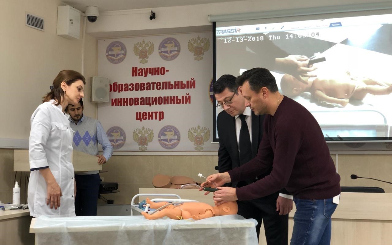
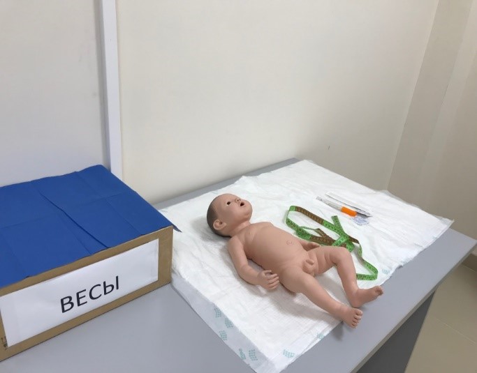
Neonatology
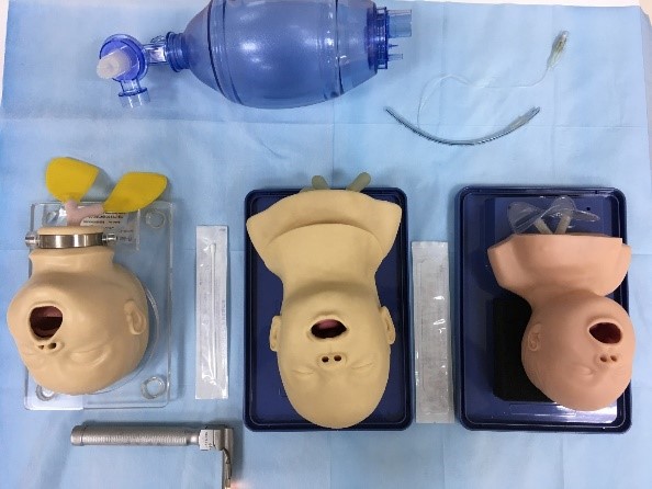 The exact anatomical structure of the simulator allows you to practice intubation of the trachea and suction of the contents of the respiratory tract.
Features:
– Realistic newborn head
– The standard position for intubation is the head thrown back
– The anatomical structure includes: nostrils, tongue, nasal cavity, pharynx, larynx, epiglottis, trachea.
– Practice intubation through the mouth and nose
– The back side can be raised to see the inner structure
– After pumping air into the tube, the lungs expand if the intubation is done correctly
– The simulator is fixed on the stand

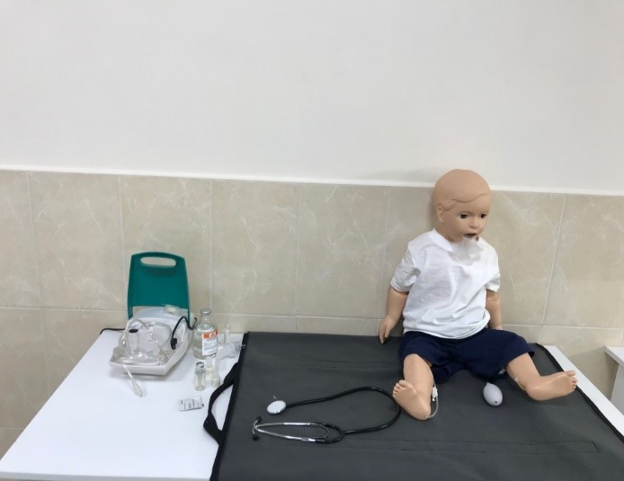
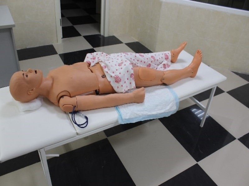
Pediatrics
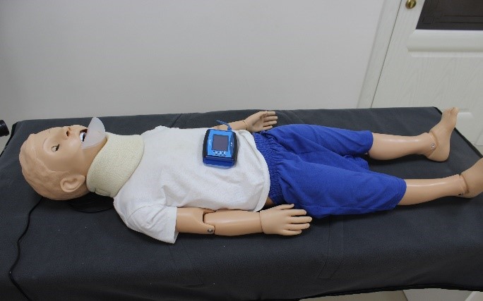 Educational resuscitation simulator for a one-year-old infant with electronic control and a computer program
- Blocking the airways when the head is tilted forward
• separate points of arterial pulse
• articulated head and jaw with tongue movable at the waist
• chest with lung and heart, soft skin
• ophthalmological procedures (eyes open/close)
• one normal eye, one dilated eye
• CPR, ALV, device for electronic monitoring of the chest compression depth and ventilation
• computer program for displaying parametric data and curves in real time on the user’s monitor
• realistic anatomical markers, eyes, fingers, hands
• intramuscular injections

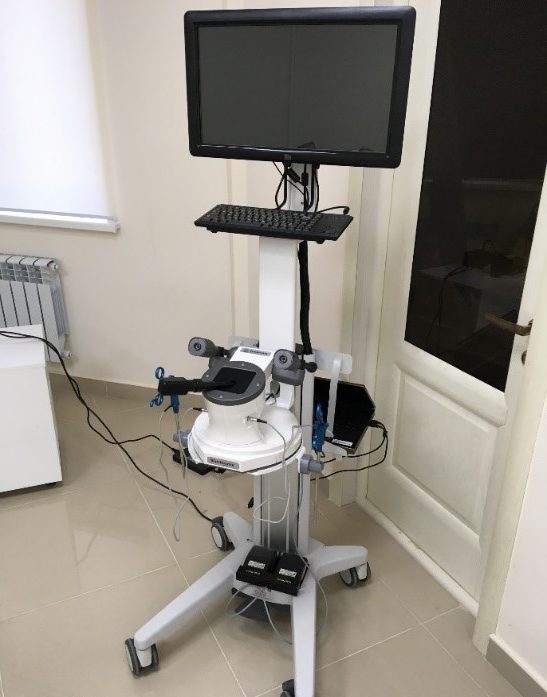
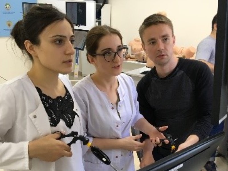
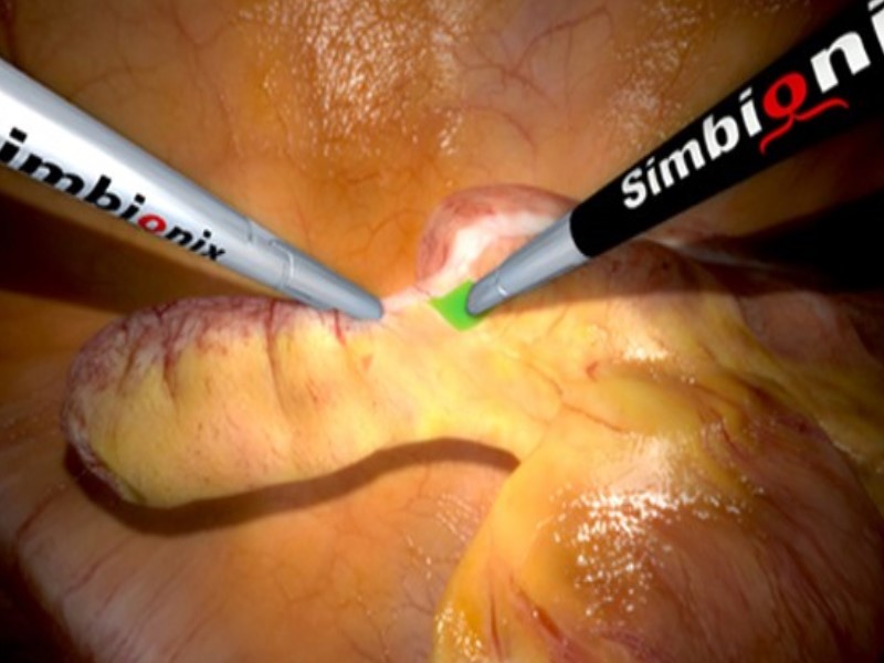
A simulator for training endoscopic surgery and practicing the technique of performing laparoscopic operations LAP Mentor.
SPECIFICATIONS
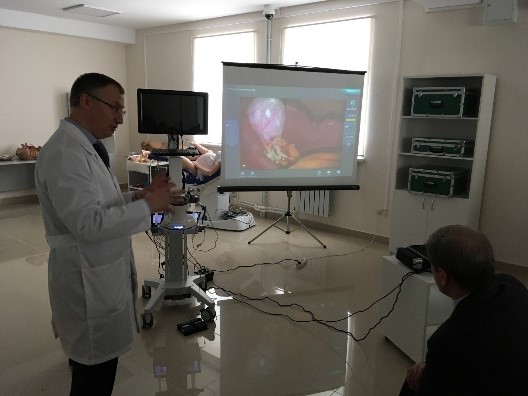
Procedural tasks
• tasks with interactive control with different levels of difficulty allow you to master the most important skills in the field of laparoscopy
Clinical procedures in full
• complex clinical scenarios with real-time simulations of complications and damages provide an opportunity to gain experience of different approaches to the procedure
Realistic learning environment
• a combination of realistic tactile feedback, detailed anatomy, realistic visualization, a wide range of surgical instruments, and a variety of trocar position configurations.
• educational content
• didactic features and tutorials such as 3D anatomical models, step-by-step real-life videos, interactive instructions, and ready-to-use courses
Assessment of the achieved level
• Comprehensive objective reports and fitness settings tools to effectively track progress in the learning process
Detachment training
• it is provided by combining LAP Mentor Express with Lap Mentor, which allows you to master the skills of an assistant, train non-technical skills and conduct training in a team.
Basic skills training modules
•Basic skills in laparoscopy
- Basic suturing skills
- Advanced suturing skills
Gynecological surgery
• Hysterectomy
• Vaginal cuff closure
• Essential skills in gynecology
General surgery
• Procedural tasks
• Laparoscopic cholecystectomy
• Cholangiography
• Appendectomy
• Incisional hernia
Thoracic surgery
• Lobectomy
Bariatric surgery
• Gastric bypass
Colorectal surgery
• Sigmoid colectomy
Urological surgery
• Nephrectomy

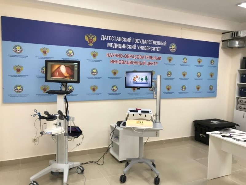


Laparoscopic training system
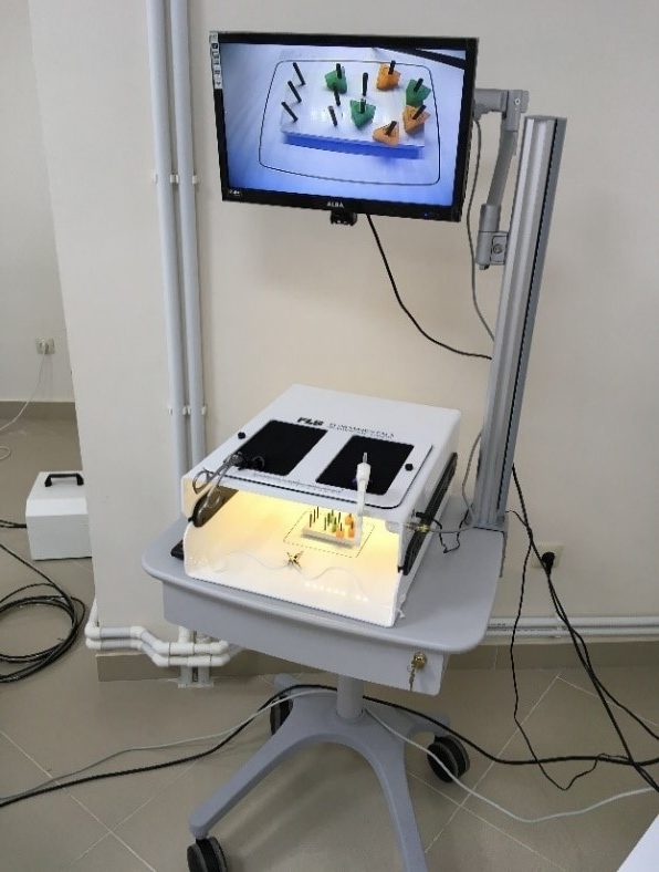 The training system includes all the functions of a simulator device, and also includes a 19-inch TV screen and a mobile cart.
Tasks:
— Pin profile
— Accurate cutting
— Ligature loop
— Knitting knots
Features:
— Leather made of durable neoprene
—Trocar pores
— USB camera in the set
—19-inch TV screen
— Mobile cart
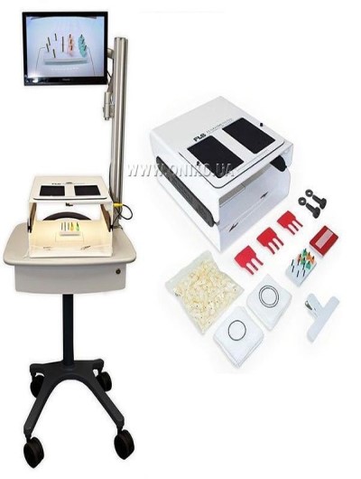
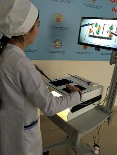
Laparoscopic surgery simulator ENSIM-S. LPR. 01

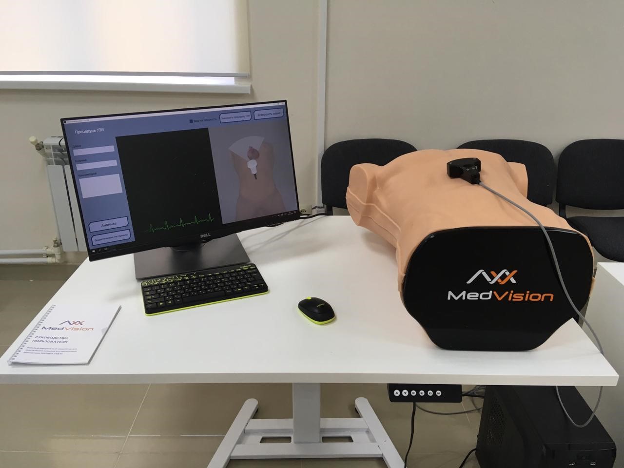
Ultrasound practical skills drilling simulator ENSIM-B.UZD.01
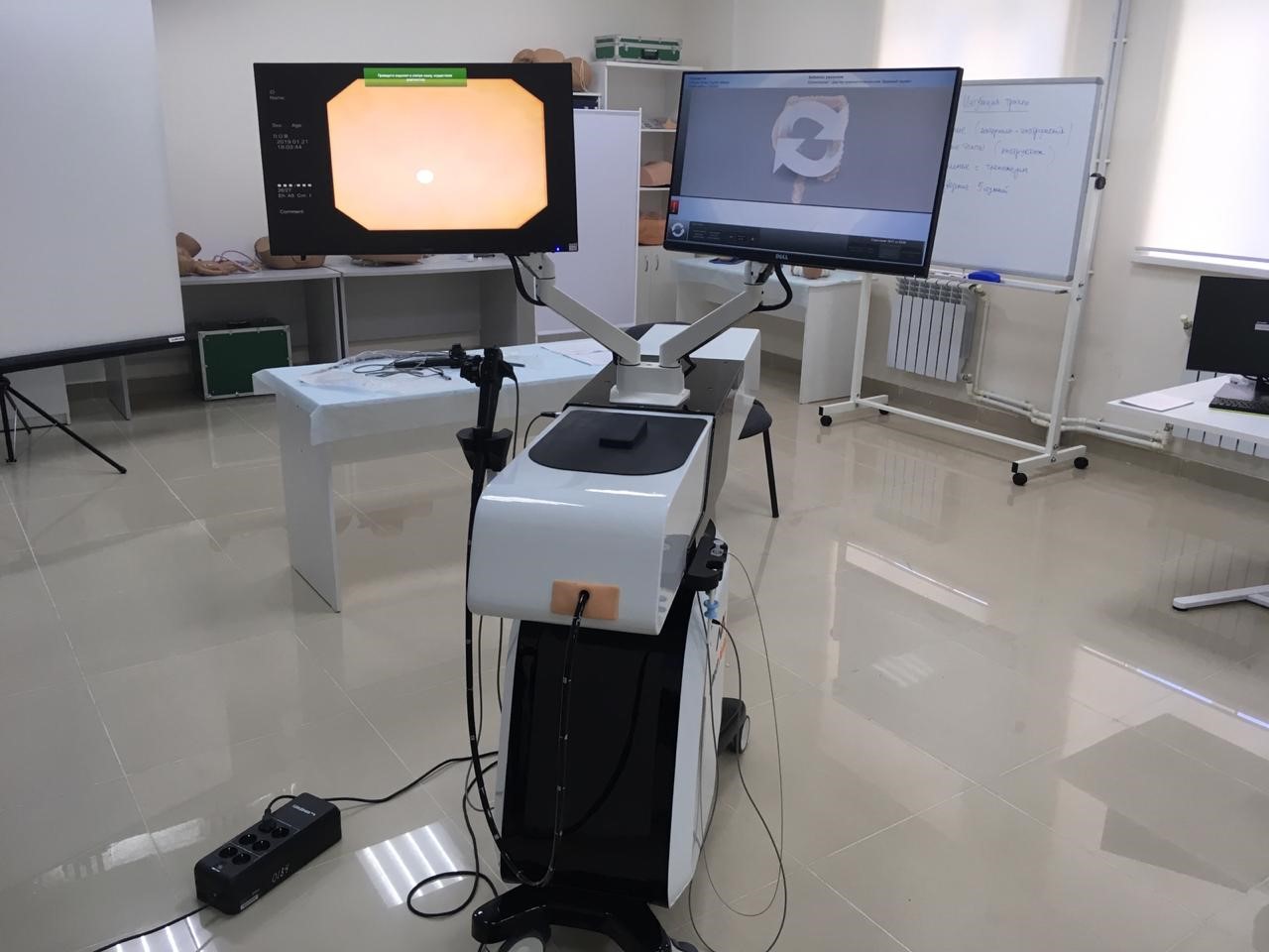
Endoscopic interventions simulator ENSIM -S.GAS.01
Interactive system of medical judgment training
Abdominal palpation skills training. LIVEPALP
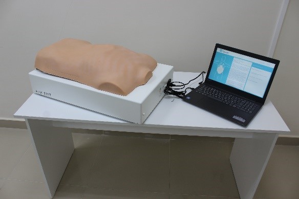 Features
•Testing and training mode.
• Visual, audible, and tactile feedback.
• Diagnosis of the most frequent pathologies of abdominal and pelvic organs.
• Differential diagnosis.
• The possibility of using it as a hybrid technique at the OSCE.
• Palpation visualization on the screen (too deep, not deep enough, or deep enough).
• Imitation of the soreness of certain areas of the abdominal cavity, muscular guarding (defense), filled and emptied state of the bladder, organomegaly of a number of organs.
• Objective assessment based on a number of criteria, such as the accuracy of the diagnosis, the completeness of the study of all the necessary areas of the abdominal wall, the proper depth of palpation, the degree of effort applied.
The LivePalp simulator, depending on the selected pathology and the training task, can simulate a palpatory picture of a number of internal organs in normal and pathological conditions, and the program keeps a protocol of the completeness of the cadet’s research, in particular, whether the following organs and areas of the abdominal wall were palpated:
• liver
• gallbladder
• stomach, epigastric area
• pancreas
• spleen
• thick intestine
• appendix
• left and right ovaries
• urinary bladder in filled and emptied states
Cholangiolithiasis
• pancreatitis
• cholecystitis
• small intestinal obstruction
• appendicitis
• diverticulitis
• acute enteritis
- hepatomegalia
- splenomegaly
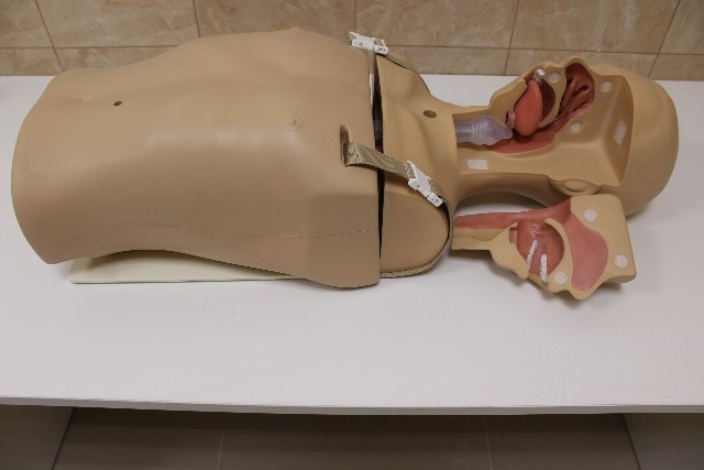
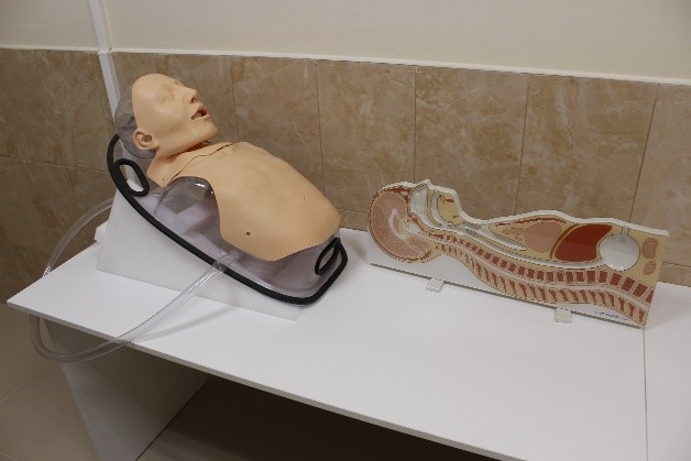
Adult dummy simulator to practice the techniques of removing foreign body from the upper respiratory tract
Realistic natural-size adult exercise dummy. The anatomically correct chest and other landmarks necessary for the correct execution of procedures are presented. Comes complete with “foreign bodies”.

Lumbar Puncture Simulator II

Skills
The lumbar puncture simulator was designed by experts in the field of medical education in order to make the development of lumbar puncture skills and their assessment more effective. The simulator allows students and specialists to exercise frequently and achieve a high level of skill in performing this procedure without putting patients at risk.
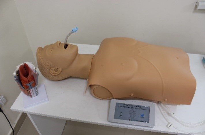
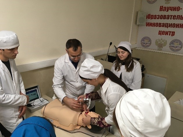
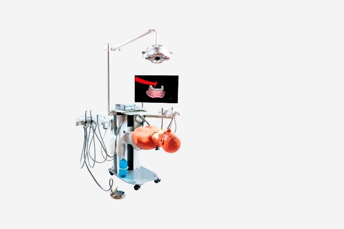
Intubation training model with control through a tablet computer.

SKILLS
• intubation
SPECIFICATIONS
• the simulator is a model of an adult’s torso
• the simulator allows you to perform manipulations:
— with a tilt of the head-chin and extension of the lower jaw
— using a sniffing position
— using a laryngoscope for intubation
— with air ducts introduction through the mouth and through the nose
— with the use of an intubation tube, laryngeal mask, double-lumen tube
- during ventilation, a chest excursion is observed
• with excessive use of the laryngoscope, the simulator emits sound signals:
—you can track the depth of intubation on the tablet
(deep/moderate/shallow)
— oesophagus intubation
— air inhaled volume per breath cycle and gastric dilatation
— determine the method of throwing the head-chin, pronging the lower jaw, sniffing position and seeing their condition on the screen
• the simulator has a touch screen with a color display, allows you to save all the results, has wireless control and the possibility of power supply from the battery and from the mains (220 V)
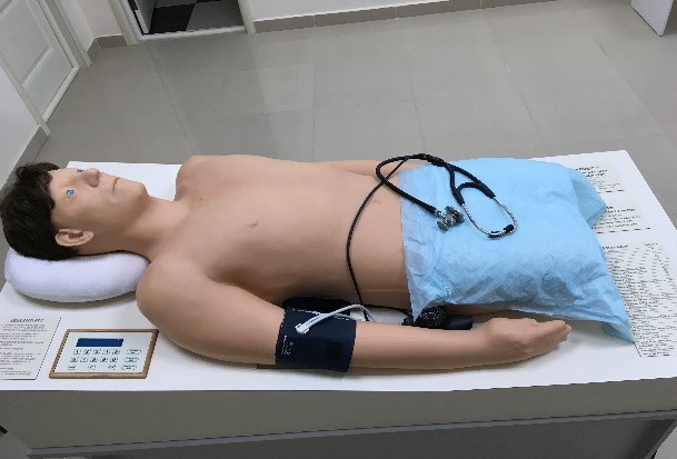
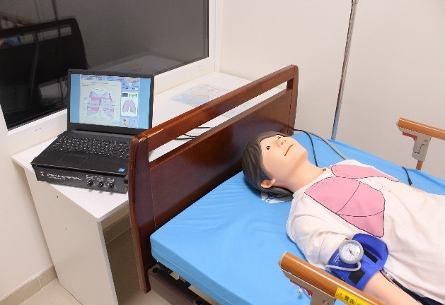
Physical examination simulator.
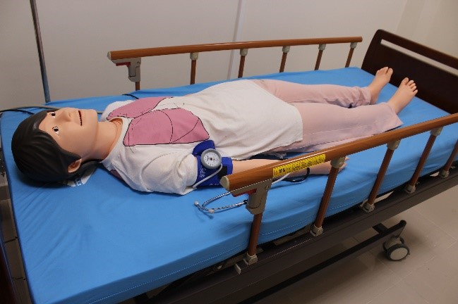 The simulator features:
Checking the pupil reflex,
Measuring blood pressure,
Determining the pulse (carotid artery / radiation artery),
Listening to breathing,
Listening to the heart, 17 sounds,
Listening to bowel sounds,
ECG imaging training. Physical inspection;
Auscultation
Simulator for aspiration and probe feeding skills
 SKILLS
• carrying aspiration catheters through the nasal cavity, mouth and tracheostomy
• aspirational manipulation
• feeding through the nasogastric probe
• manipulation of gastrotomy care
SPECIFICATIONS
• the area of the face on the middle line can be divided into two halves, which allows to confirm the real position of the injected aspiration catheter or nasogastric probe
• the model design provides an opportunity to study the anatomical structures of the nasal, oral and neck
• simulator has simulated “sputum,” which can be introduced into the nasal cavity, oral cavity or trachea using water, the viscosity of simulated “sputum” can be controlled
• it is possible to develop skills in introducing a feeding probe all the way to the bottom of the stomach
• if the probe is inserted correctly, you can hear the sounds of air bubbles and aspiration of the gastric contents
• components for probe feeding come together with an internal reservoir, and you can also use equivalent real nutrient solutions
• drain function is provided in the tank
• model is disassembled, which makes it easy to carry out its technical processing
• liquid tanks are washable
• probe aspiration (aspiration through a cannula for tracheostomy)
• feeding probe introduction all the way to the bottom of the stomach
• probe feeding (confirmation of the probe introduction correctness)
• probe treatment for gastrotomy

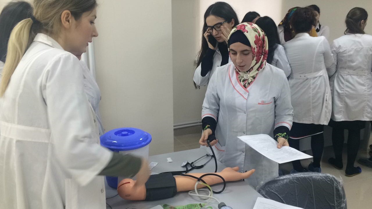
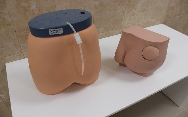
Arm simulator to measure blood pressure
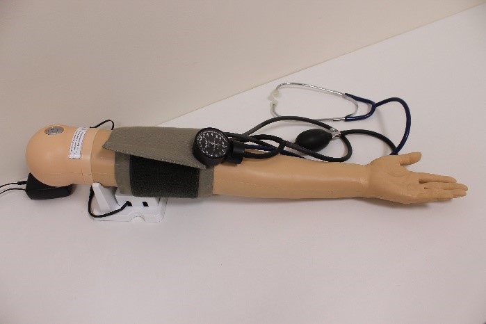 Skills
- pulse palpation on the radial and brachial arteries
- listening to the Korotkov tones of the brachial artery with a stethoscope
• systolic and diastolic pressure measurement
SPECIFICATIONS
• ability to adjust systolic/diastolic blood pressure and heart rate
• the ability to set up an ausculative gap
• displaying a graph of changes in pressure in the cuff in real time
• real-time decompression rate
• the arm skin is made of a soft material, similar to the sensations of real human skin
• touch screen with a friendly user interface 7
• the test outcomes can be saved on a USB device.
SIMULTANEOUS AUSCULTATION BY STUDENTS
• students can listen to Korotkov’s tones through the speaker
• it is possible to adjust the volume of Korotkov tones.
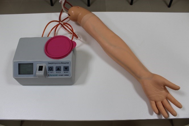
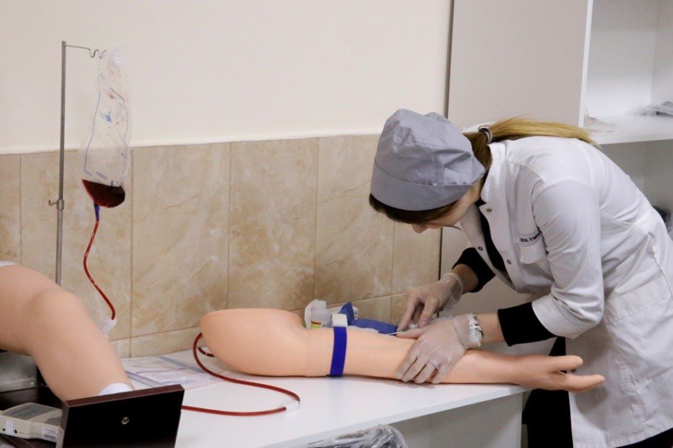
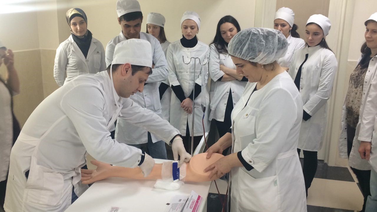
Simulator for practicing intravenous injection skills
 SKILLS
• injections into the medial saphenous vein of the arm
• injections into the lateral saphenous vein of the arm
• injections into the median cubital vein
SPECIFICATIONS
• the skin and veins resemble natural ones in their properties
- the skin pad comes with the outer covering layer, subcutaneous fat and veins
• the skin pad is easy to replace: just insert it into the gutter on the hand arm simulator
• the speed of stimulated blood flow in the arm resembles the speed of natural blood flow.
• you can use conventional or rechargeable batteries as a power source
• the simulator works in silent mode
• the single method of connecting makes it easier to connect and to separate the tube

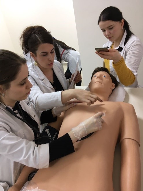
Intramuscular injections simulator
Using the simulator, you can learn a number of procedures, including the introduction of an injection needle and the injection of drug solutions.
SKILLS
• injection needle insertion
• injection of drug solutions
SPECIFICATIONS
• realistic model for intramuscular injection training
- the muscles and skeleton texture similarity of the simulator with the real ones helps to choose the right injection site. If the injection area is selected incorrectly, an audible signal appears
- the feeling that occurs when the needle is inserted is very realistic. This reduces the feeling of fear in students in real clinical work
- various procedures, such as drug infusions, can also be simulated
- injection sites and model skin are easily replaced
Simulator for teaching medical manipulations.
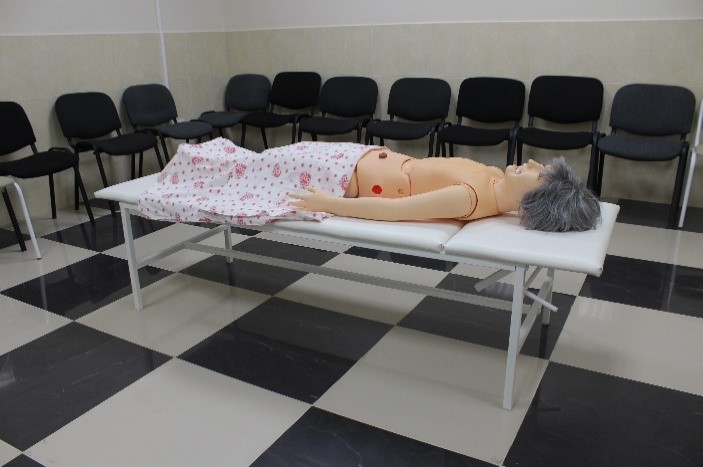
This full-length mannequin with movable joints, soft fingers and palms, the back of the head and limbs can take a natural position.
Movable jaw with tongue, removable dentition. Replaceable external genitalia (male-female), which allows you to teach catheterization techniques for both men and women. Complete set with pharyngostomy, rhinostomy, antrodrenage, tracheostomy, gastrostomy, as well as colostomy (on the transverse colon), ileostomy and suprapubic stoma.
Soft skin, mobile feet and toes, as well as a wig make this simulator very real and offer a variety of procedures and practices for patient care:
1. hair care and surgical bandages;
2. 2. taking baths and applying bandages;
3. 3. oral and dental hygiene;
4. 4. oculomotor treatment;
5. 5. intramuscular injections (arm and buttock);
6. 6. ostomy;
7. 7. naso-gastric lavage and probe feeding;
8. 8. catheterization;
9. 9. enema procedures;
10. 10. vaginal douching and taking a swab.

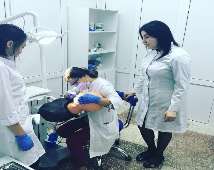
Dental chair simulator model “Superior”.
A model for practicing practical skills with periodontal pathology and teeth affected by caries and tartar
Set
- self-draining dummy with retractable articulator (0954)
- water treatment system
* surgical vacuum container on the assistant’s side
* * large tool bar with lower feed on the bracket, equipped with a booster and two basic tools
* aspiration system and water bottle, accessible from the outside
* pedal for controlling tools
* a backup pedal for installation-based control or a manual switch on the console to adjust the position of the dummy’s torso
Chest Cavity Drainage and Pneumothorax Dummy.

The dummy is designed specifically for practicing the skills of draining the chest cavity and pneumothorax in the pre-hospital period.
On the right side of the dummy, there are two skinless areas where the ribs and lungs are visible
On the left side is the area of the pneumothorax (under pressure), which allows you to vent the air accumulated in the pleural cavity, and limits the inflation of the lungs.
There is also an area for removing fluid and air from the pleural cavity by means of a tubular drainage introduced by piercing the chest wall with a trocar.
The color, volume and viscosity of the liquid to be removed is controlled by the instructor
Chest drainage; Surgical skills